RIFE CRANE Rife machines Culturization of Virus |
|
|
|
|
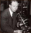
Culturization of Virus Royal R. Rife July 14, 1958
For many years scientists have desired to culture micro-organism in their filterable state. This was first accomoplished in November of 1931 when Dr. Arthur I. Kendall, Ph.D., Director of Medical Research, Northwestern University Medical School succeeded in creating a media which was called "K" media. "K" media has been demonstrated to our entire satisfaction and has the faculties or transforming non-filterable pathogenic micro-organisms into a state or transition. This would be commonly represented by a hen laying an egg whereas in this instance, the bacteria sheds a chemica1 particle which we know as a virus. Virus carry one prime inherent factor; if the bacteria is motile, the virus is motile and visa versa. The scientist will have extrema difficulty in following the virus culturization technique. The first step in growing virus micro-organisms is to take distilled water and make a triple distillation as free as possible from chlorine and flourine The Tyrode solution follows: (1 liter of triple distilled water add-0.05 grams of Na2HPO + 8.00 grams of NaCl + 0.20 grams of KCl + 1 + 0.10 grams of CaCl2) All apparatus must be sterilized before use. The air factor must be reduced as much as possible. Most virus forms become proteophilic after being in "K" media. The filterable virus forms have demonstrated to be the causitive agents of disease the theoretical and applied biology cannot be denied. Standard Berkafeld "W" and "N" filters are commonly used which are similar to Dresden china unglazed and the filtrate is usually drawn through with the aid of 2 inch water vacuum. "K" medium is fundamentally made of swine intestine. This material is relieved of all fatty substances, boiled, and the residue and fat extracted with alcohol. This requires at least five extractions with alcohol solution. Then the material is dehydrated in a dehydrating oven. If this technique is properly followed, we have from ten gallons of pig gut from the slaughter house, one pound of dessicate. A combination of two grams of the dessicate and ten cubic centimeters of Tyrode solution is placed in a test tube accurately weighed. This procedure is duplicated as many times as necessary depending on the contemplated tests. The test tubes are the placed on a rack in an autoclave for three hours. The pressure is increased very slowly to 15 psi for 1 and ½ hours and then slowness complied with and if not, the solution will be cloudy. Never more than two standard loops of the inoclaum is used which is incubated at 37 ½ deg. C for 24 hours. It is because of the difficulty of this procedure and the lack of specialized Rife virus microscope that observers have not been able to see the true filterable forms of virus, bacteria, and fungi. The electron microscope will not show these filterable forms as live entities because as all scientist know, that when the micro-organism are prepared for observation, gelatin slides, and various stains are used which combine to show obscure forms. These forms may be in the shape of round balls which is merely the grannulation of the stains or in some cases part of the micro-organism may be determined as an opaque object without detail. We have in many instances merely taken the gelatin slide and stained it without the filtrate and have nearly always attained the results. It has been an undisputed fact over many years that these virus could not be seen with any standard research microscope. This is decidedly true owing to two factors; (1) the lack of magnification, (2) improper illumination within the optical system The Rife patented microscope lamps have greatly improved the virus microscopes. We can make a positive diagnosis of any disease from which the filtered virus form can be observed under the Rife microscopes. The different virus mentioned in other reports have been checked 181 times against a color dictionary to see if the color of the various forms would chance a shade or two but they have been found to be very constant. The time for this determination varies from five minutes to one-half hour to focus in the slide and observe the true color and form of the virus in question,
SUMMARY OF PROCEDURE 1. Make media 2. Transplant block 8 mm square of un-ulcerated malignant tissue into 10 cc of Tyrode solution + 2 grams of "K" media in test tube. 3. Excite under argon filled loop at 5000 volts for 24 hours. 4. Incubate at 37 ½ deg.. C in a 2" water bath for 24 hours. We have found that by transplanting the initial culture through ten transplants from "K" media to "K" media solution (with Tyrode) with an elapsed period of 24 hours, the virulence of the inocleum is increased. Each transplant is incubated for 24 hours. Use one loop per transplant. 5. Cancer virus or "BX" will grow as a cloudiness in the solution. 6. Examine the slide under the Rife microscopes and at 15,000x, - a purplish red (cancer virus) virus will be seen. 7. Inject this virus into the mammary gland of
a rat no less than 40 rum under the epidermis and the cancer tumor will
crow.
|
" Unless we put medical freedom into the Constitution,
the time will come when medicine will organize into an undercover dictatorship
to restrict the art of healing to one class of men and deny equal privileges
to others: The Constitution of this Republic should make a special privilege
for medical freedom as well as religious freedom." -- Dr. Benjamin Rush,
signer of the Declaration of Independence


