Is
Cancer Contagious?
|
Is
Cancer Contagious?
| |
|
Rife machines By Alan Cantwell, M.D. Jackie Kennedy's rapid death from "Non-Hodgkin's lymphoma" cancer in May I994 shocked and saddened the world. Now officials of the American Cancer Society are hinting that some lymphomas may be caused by viruses. If so, are these viruses contagious? And how can we protect ourselves from them? What do we really know about the origin and cause of cancer? Are these new cancer viruses related to cancer-causing retroviruses like HIV (the AIDS virus)? As a rich woman, Mrs. Kennedy-Onassis received the best treatment for her disease at one of the most prestigious cancer hospitals in New York. Yet, within 5 months of the discovery of her lymphoma, she was dead. Even in my most paranoid state I cannot conceive of any sinister aspect of her demise. However, it is a well kept secret in medical circles that the required treatment for cancer, particularly chemotherapy and radiation, can sometimes hasten the death of cancer patients. For example, one of my patients had "routine" treatment for Hodgkin's disease, which consisted of extensive surgery followed by radiation to the chest and abdomen. After he re covered from this, he was given an intensive course of chemotherapy. Within a year of his diagnosis, and less than four months after chemotherapy, he died. At autopsy, there was no evidence of cancer. His death was caused by fatal damage to his heart and lungs by the radiation treatment he received. How much of Jackie's death can be attributed to the treatment she underwent for cancer will never be known. Cancer Mysteries Cancer kills more American women than any other disease. Cancer of the breast is the No1 killer in Anglo, Black, Latina, Chinese and Japanese-American women. No one knows the reason for this. Jackie's cancer, Non-Hodgkin's lymphoma, is among the top 10 most common forms of cancer in all races, except for Blacks. Again, no one knows why. Why do Chinese-American men have a high risk of liver cancer, when the disease is decidedly uncommon in other Americans? Why is the prostate cancer rate among Blacks in Los Angeles county 60% higher than in Anglos; and 80% higher than Asians? Why do Anglo men have a bladder cancer rate twice that of other groups? No one has an adequate explanation for these cancer occurrences. Lymphoma Cancer There are three major types of lymphoma cancer: Hodgkin's disease, Non-Hodgkin's lymphoma, and a type of lymphoma which affects the skin called mycosis fungoides. All lymphoma patients may experience similar symptoms. The exact type of lymphoma requires diagnosis by a pathologist, and depends on the microscopic appearance of the cancerous cells and tissue. The "classification" of lymphoma types is in constant flux and reappraisal. Strangely, the cancer cells of Jackie's Non-Hodgkin's lymphoma were reported as a new type called "Ki-1." Hodgkin's disease is one of the very few forms of cancer that has long been considered as having a possible infectious origin. For decades, epidemiologists have recognized "cluster cases' of Hodgkin's. The actual number of cases was so high in certain communities that it was highly unlikely to be due to statistical chance. Even so, definite proof of a Hodgkin's disease agent has not been forthcoming. Some scientists are now suspecting a possible cancer causing retrovirus. The Cancer Microbe All this would not surprise my mentor, the late Virginia Livingston MD, who for 40 years claimed that various bacterial and viral forms of "the cancer microbe" were responsible for the infectiousness and possible contagiosity of cancer. Long before the discovery of the AIDS virus, she stressed care in the sexual arena, as well as the importance of hygiene, diet and supplements in the treatment of cancer. Her use of vaccines made from the patient's own cancer bacteria ultimately resulted in the condemnation of her cancer research and her treatment methods for cancer. Robert Gallo MD, the co-discoverer of the AIDS virus, denounced her scientific work as insanity. Over the past century phvsicians have refused to believe that an infectious agent could cause cancer. In the 1970s, scientists proved that animal cancer viruses were capable of causing cancer, but there was no proof that viruses caused human cancer. Now with the discovery of cancer-causing retroviruses, such as the AIDS virus, doctors are reconsidering the idea. The history of modem medicine seems to prove Cantwell's law that "Most physicians are wrong in their understanding of most diseases, most of the time." Why is Non-Hodgkin's lymphoma one of the fastest rising cancers in the U.S.? Is the rise related to drug use (legal and illegal), immunosuppression from environmental sources, or radiation effects? No one knows. My own published research into the cause of all three types of lymphoma indicates that "cancer microbes" are the infectious agent. My studies confirm Livingston's research, as well as the cancer microbe research of dozens of other scientists throughout the world who have implicated bacteria in various forms of cancer. This work is documented in my book The Cancer Microbe (1990). Over thirty of my published papers on AIDS, cancer, and other immunologic diseases, are available in medical libraries. And anyone hooked into a computer network can get abstracts of these papers through "Medlars 11," the National Library of Medicine's computer retrieval service. The startling cancer microbe discoveries are ignored by the cancer establishment. Undoubtedly the recognition of a heretofore unnoticed infectious agent in cancer and AIDS would provoke havoc in medical science. Thus, cancer bacteria remain "in the closet" and the research "politically incorrect." Death by Cancer So many people have been done in by cancer at a convenient time in history that conspiracy buffs wonder if you can "give" people cancer. I recall the propitious cancer deaths of Jack Ruby, William Casey, Martha Mitchell, the Shah of Iran, Mae Brussell, and others. (Even O.J. Simpson was tested for lymphoma cancer in a swollen lymph node while imprisoned awaiting trial for murder.) Can you kill people by injecting them with cancer viruses and bacteria? Of course! No one in their right mind would want blood from a dying cancer or AIDS patient. Can you give a person cancer? If cancer in animals can be caused by injecting them with cancer viruses and bacteria, it would certainly be possible to do the same with human beings! Cancer: One Disease or Many? The American Cancer Society constantlv reminds us that there are many "different" kinds of cancer. This concept is highly effective in raising money for each type of cancer that the ACS promotes. But in reality there is a close relationship between certain forms of cancer, particularly lymphoma and leukemia (cancer of the blood). In the 1970s, virologists discovered that animal retrovirus infection (similar to the AIDS virus) could cause an increase in lymphoma and leukemia in the animals, as well as immunosuppression. In 1983, a year before the official discovery of HIV, Harvard veterinarian Myron Essex found that 25% of gay AIDS patients had viral antibodies to blood from a case of "human T-cell leukemia," caused by a new retrovirus called "human T-cell leukemia virus-1." When Robert Gallo announced his discovery of the AIDS virus in 1984, he believed the virus was related to this new family of leukemia viruses. Thus, he first named the AIDS virus "Human T-cell leukemia virus-3." Later, the virus was renamed HIV: the human immunodeficiency virus. When the AIDS virus (HIV) was introduced into the gay community in the late 1970s, the incidence of Kaposi's sarcoma (a previously rare form of cancer), as well as the incidence of Non-Hodgkin's disease, began to skyrocket in HIV-infected gay men. Both types of cancer had previously been associated with immunodepression. For example, in the mid-1970s the normal incidence of Kaposi's sarcoma increased 400 to 500 times in transplant patients who were routinely immunosuppressed with prescribed drugs as part of the procedure. In 1981, the year the AIDS epidemic became official, Bijan Safai and Robert Good, scientists at Memorial Sloan Kettering Cancer Center in New York City, declared a close association between Kaposi's sarcoma, leukemia and lymphoma. They proposed all three cancers be considered "as part of a spectrum of disease affecting the lymphoreticular system." The American Cancer Society is unlikely to remind its contributors that treatment for one form of cancer can cause the formation of another "different" cancer. For instance, after chemotherapy about three percent of Hodgkin's disease patients go on to develop Non-Hodgkin's lymphoma, or leukemia. Officially, the reason for these "second cancers" is unknown. Surprisingly, most cancer experts assume two different diseases are involved. They theorize that chemotherapeutic drugs depress the immune system, resulting in the development of "new" cancers. Is Cancer Contagious? Physicians now understand that sexually transmitted retroviruses, such as HIV, can cause cancer and immunosuppression. Nevertheless, doctors continue to tell patients (unless they are gay) that cancer is neither infectious or contagious. What about the transmissability of new retroviruses that have been identified in certain human leukemia cases? So far, the cancer establishment is not commenting. Do cancer viruses and the increasing rates of certain forms of cancer, such as Non-Hodgkin's lymphoma, have a relationship to the AIDS epidemic? In the absence of a positive HIV test, I would say no. (Both Jackie Kennedy and O.J. Simpson tested negative for HIV.) In my book, Queer Blood: The Secret AIDS Genocide Plot (1993), I describe the genetic engineering of animal cancer viruses in the early 1970s that, in my opinion, resulted in the creation the AIDS virus. (Are there any PARANOIA readers who actually believe AIDS comes from African green monkeys?) Man-Made Viruses Whether one agrees with the idea that AIDS is a man-made epidemic, it is a fact that we now deal with viruses that are "natural" (so-called endogenous viruses); and viruses that are genetically manipulated, man-made, and "unnatural" (exogenous viruses). These laboratory-created viruses are now used as an integral part of the new gene splicing technology. Engineered and altered animal cancer viruses, such as the mouse leukemia virus, have already been used in human genetic experiments. If cancer viruses can be injected into people to make them well, they certainly can be injected into people to make them sick! Can genetically engineered viruses be covertly seeded into select populations for political and genocidal purposes? Was the "introduction" of the AIDS virus into the Black African population, and into the U.S. gay male population, a deliberate attempt to eliminate these groups? Govemment AIDS experts repeatedly tell us that AIDS began as a Black African disease when an African green monkey "jumped species." The U.S. public was initially told AIDS is a "gay" disease, caused by anal sex and drugs. But who can conceive of a Black heterosexual epidemic in Africa that transformed itself into an exclusively young white gay male epidemic in New York, Los Angeles, and San Francisco? Recently a physician colleague referred a patient to me with a written diagnosis of "AIDS caused by homsexuality." This indicates how powerful and effective the government’s disinformation program has been in convincing the public (and some physicians) that AIDS is a gay disease. Now that 4 million men and women are infected with HIV worldwide, do people really think most were sodomized? Coping with Cancer Jacqueline Kennedy-Onassis's death from cancer, despite access to the best treatments and doctors that money could buy, certainly justifies a degree of cancerophobia. Her rapid demise makes us wonder if her treatment killed her quicker that her lymphoma. (Some lymphoma patients live for many years.) But did she have any treatment options? If you had cancer, would you go against the advice and wisdom of a cancer expert? Who would turn down chemotherapy and radiation if they thought it would save their life? How do doctors separate the toxic effects of cancer from the toxic effects of treatment with chemotherapeutic drugs, surgery, radiation and sterids? Will doctors be able to save us if we are diagnosed with cancer? Can we catch cancer-causing viruses from our family, our friends, our lovers? Is this reason for paranoia? You bet it is!" Doctor Cantwell is the author of The Cancer Microbe, Queer Blood: The Secret AIDSGenocide Plot ($12.95), and AIDS: The Mystery & The Solution ($9.95). All books are available from Aries Rising Press, P0 Box 29532, Los Angeles, CA 90029. (213) 462 6458. Enclose $2.00 s&h for one book; $3.00 s&h for two or more. CA residents must add sales tax. Source: Paranoia, winter 1994/95
THE ROLE OF VIRUSES Objective: The students will learn how viruses contribute to the development
of Cancer. Background: Viruses are a unique group of organisms that grow only in
the cells of bacteria, fungi ,protists, plants and animals. A typical
virus is made of a protein shell or a capsid , which surrounds a nucleic
acid core. The genome may be DNA or RNA. The function of the capsid is
to protect the viral genome while the virus is outside of living host
cells. They range in size between 10 to 200 microns. In order to replicate
they infect and take over the host cell. Whole viruses never arise directly from preexisting viruses.
They develop and reproduce only within cells of specific hosts. Since
viruses do not have many of the necessary structures like ribosomes to
produce proteins, they must redirect the genome of the host cell in order
to reproduce themselves. Viruses Examples of Human Cancer Viruses
Researchers have identified the role of a protein segment that allows
some cancer-causing viruses to latch onto and infect cells. This may lead to new therapies that target these viruses. Viruses, Variations and Environment-Studies on For many years the human papillomavirus (HPV) has been suspected as
a possible culprit in the etiology of oral cancer. Two types of the virus,
HPV-16 and HPV-18, are found in oral cancer tissues more frequently than
in normal tissues. Scientists are still not sure, though, how HPV might
contribute to the development of oral cancer. Viruses
and Cancer, 1962: What They Knew Yes,
Virginia, Viruses Do Cause Cancer Patrick S. Moore, MD, MPH, Director
http://www.geocities.com/abettica/cancervirus.html . Cancer is a disease of aging. It is caused
by heritable changes in the cell's genetic material. Cancer cells divide
to produce daughter cancer cells. Much of what we now know about the
genes involved in the development of cancer is attributable to research
into RNA and DNA tumor viruses. There are 7 families of viruses associated
with tumors (1 RNA and 6 DNA families). The DNA viruses include the
hepadnaviruses, the polyomavirus, the papillomaviruses, the adenoviruses,
the herpesviruses and the poxviruses. The RNA virus family is the retrovivuses
(sub group oncoviruses). It is important to note that in humans the
association of viruses and cancer is not causal and is often correlative
or ambiguous. At best viruses are thought to be cofactors or co-carcinogens
in the development of human tumors. This underscores the fact that
cancer is a multistep process. It is the relative simplicity of the
viral genome (compared to the enormous complexity of the cellular genetic
material) that has permitted the identification of genes involved in
the genesis of human cancer. SV-40, a pro-cancer virus in vaccines In 1955, Jonas Salk performed a medical miracle when he discovered how to mass produce polio vaccine by growing it on the kidneys of rhesus monkeys. While there is no question that thousands were saved from the ravages of polio by the Salk vaccine, by 1960 a problem had surfaced -- researchers had isolated a viral contaminate in the vaccine, Simian (monkey) Virus # 40. It seems that when the live polio virus grown on monkey tissues was extracted for vaccine production this SV-40 virus was extracted as well. When SV-40 was injected into research animals it produced brain cancer. It appears our government didn't wish to create a public panic or discredit the public health service, because instead of recalling the tainted vaccines, it quietly ordered the manufacturers to find a monkey free of SV-40 and continue production. As of 1963, the rhesus monkey had been replaced with the African green monkey for production of a safer polio vaccine, but between the years of 1955 and 1963 as many as 98 million Americans had received doses of live polio virus vaccines tainted with SV-40. Nowadays SV-40 has appeared in 61% of all new cancer patients -- patients too young to have received the contaminated vaccine being administered forty years ago who are now believed to have been infected by human to human transmission. Being a blood born organism, it is also suspected that SV-40 is transmissible from mother to child during pregnancy. The more this matter is researched the more startling the evidence. Senior epidemiologist at the National Institutes of Health, Dr. Howard Strickler, has plotted a geographic pattern to the cancers associated with SV-40 helping to confirm its link to the tainted vaccine. People who lived in Massachusetts and Illinois who received identified lot numbers of the contaminated vaccine administered in the 1950s are now demonstrating ten times the rate of the osteosarcoma bone tumors as those who received vaccine free of the SV-40 contaminate in other parts of the country. DNA Polyoma Viruses In 1964, studies were conducted on a polyoma virus (a tumor-producing DNA virus). It was discovered that the persistent genetic DNA material in the polyoma virus brought about malignant transformations in hamster embryo cell cultures. This was reported in the November 23, 1964 issue of the Journal of the American Medical Association. SV-40 is one example of a DNA polyoma virus. Polyoma (many tumor-causing) viruses cause prolonged infection where tissue is destroyed, integrate into the hosts genetic material, are capable of mutating a cell, may reproduce after coming into contact with a 'helper' virus, enable the separate replication of the viral genome, can generate immune responses, and they can induce malignancy. Scientists are amazed at how little genetic information these viruses carry in proportion to the damage they can cause. The 'D' in DNA and the 'R' in RNA have characteristics which are dependent on the kind of sugar molecule associated with it. DNA exists predominantly in the nucleus, but is also represented in the cytoplasm and in the mitochondria. RNA is also present in the cytoplasm. When viral RNA or DNA combines with the genetic material in the cell itself, the viral genetic material can become part of the host cell genetic code, altering the genetic structure of the cell. When the altered cell duplicates, the encoded viral genetic material may affect cellular processes in such a way as to produce abnormal cells, which sometimes become malignant or cancerous. Cancer-Causing RNA Viruses and DNA proviruses The discovery in 1975 that viruses causing cancer in animals had a special enzyme called reverse transcriptase makes the problem even more interesting. These kind of viruses are called RNA viruses. When an RNA virus has the reverse transcriptase enzyme within its structure, it allows the virus to actually form strands of DNA which easily integrate with the DNA of the host cell which it infects. Studies by Dr. Robert Simpson of Rutgers University indicate that RNA viruses which do not cause cancer can also form DNA, even without the presence of reverse transcriptase. DNA formed in this way from an RNA virus is called a provirus. It is known that some non-cancerous viruses have a tendency to exist as proviruses for long periods of time in cells without causing any apparent disease. In other words, they remain latent. Some examples of common RNA viruses that do not cause cancer, per se, but have the capacity to form proviruses are influenza, measles, mumps and polio viruses. Viruses as Catalysts for Cancer An article in the January 6, 1962 Science Newsletter indicated that 'common human viruses act as carriers in causing cancer by interacting with cancer-causing chemicals; this has been indicated by experiments which show that cancer-causing substances that are present in too small a quantity by itself will become active and create tumors when combined with single doses of virus. Malignant tumors appeared in five type of injected mice.' The viruses mentioned were ECHO9, B-4, Coxsackie, and Polio virus 2. The article further indicated that 'viruses may also activate other cancer causing substances besides chemicals in the environment, such as DMBA, AF, and DBA.' Even common non-tumor viruses, including those in smallpox vaccine and polio virus 2, can act as carcinogens. It was reported in Science on December 15, 1961 that these common viruses acted as catalysts in producing cancer when given to mice in combination with known organic carcinogens in amounts too small to induce tumors themselves. This means that some vaccinations will induce cancer, when combined with the growing problem of environmental pollution from toxic by-products of agriculture (pesticides on and in food) and industry. A Listing of Cancer Causing Microbes The July 14th 1997 issue of Business Week has an article in it about how many cancers are being linked to various viruses, and bacteria ,and parasites. Among the organisms now linked to cancer are as follows
References Fisher, B. L. (1997). Workshop on Simian Virus 40: A Possible Human Polyomavirus. National Vaccine Information Center, January 27, On-line at http://www.909shot.com/polio197.html Carbone, M., et al. (1996). SV-40 Like Sequences in Human Bone Tumors. Oncogene, 13(3), 527-535. Elswood, B. F., & Stricker, R. B. (1995). Polio Vaccines and the Origin of AIDS. Medical Hypotheses, 42(6), 347-354. Krieg, P., Amtmann E, Jonas, D., Fischer, H., Zang, K., & Sauer G. (1981). Episomal Simian Virus 40 Genomes in Human Brain Tumors. Proceedings of the National Academy of Sciences of the United States of America, 78(10), 6446-6450. Lednicky, J. A., Garcea, R. L., Bergsagel, D. J., & Butel, J. S. (1995). Natural Simian Virus 40 Strains are Present in Human choroid Plexus and Ependymoma tumors. Virology, 212(2), 710-717. Martini, F., et al. (1995). Human Brain Tumors and Simian Virus 40. Journal of the National Cancer Institute, 87(17), 1331. Martini, F., et al. (1996). SV-40 Early Region and Large T Antigen in Human Brain Tumors, Peripheral Blood Cells, and Sperm Fluids From Healthy Individuals. Cancer Research, 56(20), 4820-4825. Pass, H. I., Kennedy, R. C., & Carbone, M. (1996). Evidence for and Implications of SV-40 Like Sequences in Human Mesotheliomas. Important Advances in Oncology, 89-108. Rock, A. (1996). The Lethal Dangers of the Billion Dollar Vaccine Business. Money, December, pages 148-163. Tognon, M., et al. (1996). Large T Antigen Coding Sequences of Two DNA Tumor Viruses, BK and SV-40, and Nonrandom Chromosome Changes in Two Glioblastoma Cell Lines. Cancer Genetics and Cytogenics, 90(1), 17-23. Inherited virus may play role in breast cancer Subcellular Life Forms Indeed, besides my love of elegance and my morbid fascination with parasites, the main reason subcellular life forms appeal to me is that they challenge our naive notion of organisms as entities with clear, well-defined boundaries. It's clear by now that life doesn't respect this simple picture. Whenever a pattern of any sort, however abstract, is able to replicate itself, it does! Typically these patterns overlap and interact in subtle ways, so one can't easily say where one ends and the other begins. These are the main kinds of subcellular life forms that I know about so far: Viruses One thing to keep in mind: these life forms are small. Remember that DNA is a double helix containing information in the form of AT and CG "base pairs" (paired molecules of adenosine and thymine, or cytosine and guanine), while single-stranded RNA is a single helix containing information in the form of A, U, C, and G "bases" (molecules of adenosine, uracil, cytosine and guanine). The human genome is made of DNA and contains about 5 billion base pairs. The genome of a bacterium is also made of DNA but has less than 10 million bases. The potato spindle tuber viroid, on the other hand, is nothing but a circular loop of RNA consisting of 359 bases! Small, simple - but effective! --------------------------------------------------------------------------------
Apart from their intrinsic interest, viruses are important because they cause many diseases among humans, such as: the common cold There is standard taxonomy of viruses [CT] [Ma2], [F], but I will content myself with a rough classification into the following 3 sorts: DNA viruses Retroviruses DNA viruses Like retroviruses, some DNA viruses work their way into the nucleus of their host cell and then copy themselves into the host's DNA. An example is the hepatitus B virus, which occupies liver cells. This can cause tumors.
(Actually the smallest one, the hepatitis delta agent (HDV), is quite different from all the rest. Like a virusoid, it is a circular loop of RNA that can only reproduce in cells infected by a helper virus, the hepatitus B virus. But unlike a virusoid, it affects animals rather than plants, it has its own protein coat, and its genome is bigger than that of a virusoid, having 1,700 nucleotides instead of a mere 350 or so. However, its genome is much smaller than that of any other virus.) One can broadly classify RNA viruses into: positive-strand RNA viruses
It has been estimated that between .01% and .1% of the genome of wild and laboratory mice consists of endogenous retroviruses. The same is probably true for humans. to form protein coats - since most mammalian DNA serves no known purpose, the above figures may be drastic underestimates. Indeed, 97% of human DNA is so-called "junk" DNA of this sort! Retroviruses are important in genetic engineering because they raised for the first time the possibility that RNA could be transcribed into DNA, rather than the reverse. In fact, some of them are currently being deliberately used by scientists to add new genes to mammalian cells. In addition, retroviruses are important because AIDS is caused by a retrovirus: the human immunodeficiency virus (HIV). This is part of why AIDS is so difficult to treat. Most usual ways of killing viruses have no effect on retroviruses when they are latent in the DNA of the host cell. Many retroviruses cause tumors in animals. These viruses contain host-derived genetic information. --------------------------------------------------------------------------------
Most known viroids cause diseases in plants. The first viroid was discovered in 1971, by Diener. It's called the potato spindle tuber virus (PSTV), since it causes a disease that makes potatos abnormally long and sometimes cracked. At the time, Diener's isolation of the viroid causing this disease met with some skepticism, since it was so much smaller than any known virus. By 1991, however, at least 15 plant diseases had been traced to viroids. There are also 2 viroids known, the hop latent viroid (HLV) and a viroid living in grapevines, that cause no known symptoms! This raises the fascinating possibility that there could be more such viroids lurking around. The complete molecular structure of many viroids has been worked out, which has allowed a classification of viroids on the basis of their RNA sequences. Roughly speaking, there are a large family of viroids that share many features with PSTV, together with one viroid that seems very different: the avocado sunblotch viroid (ASBV). McInnes and Simons have proposed a further classification of the PSTV-type viroids into three kinds [Ma1]. It is clear from these RNA sequences that viroids are not "degenerate viruses", as had once been thought. They are quite different from any known viruses. One interesting theory is that they arose from RNA that escaped from cell nuclei. It's also interesting that all viroid diseases have been detected in the 20th century, some quite recently - in contrast to diseases caused by viruses. Also, many viroid diseases have been spreading after their discovery, often due to human activity. A fascinating example is the coconut cadang-cadang viroid (CCCV), a disease of coconuts which has been spreading throughout the Phillipines. On the island of Luzon, a puzzling feature of this disease was that it only affected crops owned by speakers of Bicalano, while adjacent crops owned by speakers of Tagalog went unharmed! Eventually people realized that the viroids were spread by workers cutting the palms. Tagalog owners prefer to hire Tagalog workers, while Bicalanos hire Bicalanos, some of whom came from an area where the disease was prevalent. (See the article by Maramarosch entitled "The Cadang-Cadang Viroid Disease of Palms" [D].) Because of this sort of epidemiology, Diener has suggested that viroids may be latent to their native host plants (like HLV), becoming pathogenic only when transferred to other species thanks to agriculture. Indeed, the viroid causing tomato "planta macho" disease in Mexico, TPMV, has also been found in wild plants there, where it seems sometimes "recover" from ASBV by sending up a new shoot. This new shoot is still infected with the viroid, but it shows no symptoms other than reduced fruit yield. Descendents of such a "recovered" tree are also infected with the viroid, and also symptomless, except for reduced fruit yield. Thus the avocado appears able to "come to terms" with the viroid in some way. Personally, I'd like to raise this possibility: that some viroids actually play a beneficial role in their native host plants! This may seem surprising, but when we compare the behavior of plasmids, it may seem less so. --------------------------------------------------------------------------------
In short, a virusoid is a parasite of its helper virus. But it's not always so simple. Sometimes the helper virus is unable to reproduce unless the virusoid is present! Then we have symbiosis rather than parasitism. The first virusoids were discovered in the early 1980s in Australia, associated with viruses causing plant diseases such as velevet tobacco mottle (VTMoV), solanum nodiflorum mottle (SNMV), lucerne transient streak (LTSV), and subterranean clover mottle (SCMoV). An interesting theory about the origin of virsoids is that in plants infected with both viruses and viroids, the viroids got encapsidated in the viruses and later lost their ability to reproduce independently. At this point, I should admit that the terminology concerning virusoids is quite confusing to me. People sometime use "satellite RNA" as a synonym for "virusoid", but I'm not always sure when it's supposed to be an exact synonym. Diener and Prusiner define a "satellite RNA" to be a "small RNA that does becomes packaged in protein shell made from coat proteins of another, unrelated, helper virus, on which the satellite RNA depends for its reproduction". The similar-sounding term "satellite virus" appears to be reserved for an RNA virus that depends for its reproduction on an unrelated helper virus, but whose genome codes for its own protein coat. --------------------------------------------------------------------------------
However, is difficult for me to resist the impression that plasmids are just as "alive" as viruses. Indeed, some viruses become plasmids when parts of them are missing! For example, the "lambda bacteriophage" is a virus that infects the intestinal bacterium E. coli, but "lambda dv particles", which arise from the lambda phage simply by deleting some DNA, are plasmids. The lambda phage multiplies inside its host and then kills it by "lysis", which destroys the cell membrane and releases lots of copies. The lambda dv particles, on the other hand, stays in the cell in a fairly stable number of copies and does not kill its host. The difference is that while the lambda dv particles contain genes for replication, they lack genes for lysis and the protein coat. If we think of plasmids as life forms, we must admit that they are very successful. Many plasmids spread so thoroughly in cultures of bacteria that less than one cell in 100,000 lacks a copy! Some kinds of plasmids contain genes that help make sure copies are efficiently passed on to both daughter cells when the host cell divides. F plasmids have a particularly clever mechanism - they temporarily inhibit cell division when they have not yet replicated inside the host! Plasmids are diverse and very interesting. Some important kinds are: R Plasmids Cryptic Plasmids Phasmids Some good books on plasmids include "Plasmids" by Paul Broda [B], "Bacterial Plasmids" by Kimber Hardy [H], and "Plasmids of Eukaryotes: Fundamentals and Applications" by K. Esser et al [E].
R plasmids contain genes that give their bacterial hosts resistance to antibiotics as well as to poisonous metal ions such as arsenic, silver, copper, mercury, lead, zinc and so on. Because many R plasmids are conjugative, this resistance can spread from one bacterium to another. Because they can live in more than one species of bacteria, R plasmids can also spread resistance between bacteria of different species! Spread of resistance to antibiotics is now a major problem in medicine. Drugs which were used for many years to control bacterial diseases are now becoming helpless against new resistant strains. The problem has been made worse by the tendency for doctors and veterinarians to use antibiotics when they aren't strictly necessary, for example as part of livestock food. As a result an environment is created where bacteria with resistance have a great competitive advantage, so they spread rapidly. It has also recently been found that weeds growing near crops that were genetically engineered to resist herbicides can acquire this trait. I'm not sure, but I suspect that this happens via plasmids as well. R plasmids make it clear that the idea of evolution as a battle between species with separately evolving genomes is a great oversimplification. Instead, genetic communication and cooperation between different species can be very important.
F plasmids give their hosts no known traits besides these sex pili. The evolutionary origins of sex are much debated these days; we see here the fascinating possibility that sex can originate as a kind of disease whose sole function is to spread a parasite!
In short, different strains of colicin plasmid compete with each other using the resources of their hosts. A colicin plasmid will confer an advantage to its host bacteria if the other strains of bacteria nearby do not have a colicin plasmid. However, when there are many different strains of colicin plasmid present, all strains of host bacteria suffer. Thus there is a certain similarity between colicin plasmids and "protection rackets" run by Mafia-like gangs. Colicin plasmids are not the only sort of plasmids that exhibit incompatibility. Similar plasmids tend to be incompatible with each other, while drastically different plasmids are usually compatible. One theory is that incompatible plasmids use the same mechanisms to maintain their copy number. In a cell containing two incompatible sorts of plasmid, their reproduction is blocked until the total number of copies of the two together drops to the copy number of each one. This is an unstable situation, especially for plasmids with a low copy number, so eventually descendants of the host cell contain only one or the other plasmid.
"Vibrio cholerae", the cause of cholera, is a bacterium whose genes code for a diarrhea-causing toxin. The DNA of these genes is closely related to the DNA of certain virulence plasmids infecting E. coli - so closely that there is almost certainly a common ancestor. For example, Vibrio cholerae could have evolved from an earlier bacterium by permanently integrating the DNA from a virulence plasmid into its genome. Strains of bacteria and viruses often become less virulent as they coevolve with their hosts. Thus one may wonder what evolutionary advantage a virulence plasmid could confer to the bacteria containing it. In the case of bacteria causing diarrhea, there is an obvious possibility: diarrhea can serve as a mechanism for spreading the bacteria - and their plasmids - that cause it!
It's worth noting that some of these chemicals are secreted by plants as part of a defense against bacteria. Thus we probably have a kind of natural chemical arms race going on here. Other metabolic plasmids allow bacteria to degrade herbicides like 2,4-D, as well as certain detergents! People are investigating the use of such plasmids to help biodegrade pollution. Tumor-Causing Plasmids Cryptic Plasmids
The lambda phage is a virus that specializes in invading bacteria such as E. coli. In nature, its protein coat latches onto the bacterial cell membrane and injects the phage DNA into the bacterium. Biotechnologists have taken advantage of this by using the lambda phage protein coat to inject a cosmid into the bacterium! Once inside, the cosmid replicates like a plasmid and, like a plasmid, integrates its DNA into the genome of the bacterium.
--------------------------------------------------------------------------------
In addition to transposons, there is plenty of other DNA in our chromosomes that doesn't seem to code for proteins. This is sometimes called "junk DNA". It comes in various distinct forms, such as "introns", "satellite DNA", and "pseudogenes". In fact, junk DNA makes up about 97% of the human genome! Clearly despite its derogatory name, it's worth understanding and potentially very important. However, since transposons are the most "organism-like" of junk DNA, I will only talk about them here. There is a fair amount of genetic evidence that transposons spread "horizontally" between sexually isolated species in addition to being "vertically" passed down the evolutionary tree. However, the mechanisms of this horizontal transmission are poorly understood. One interesting fact is that certain viruses, the baculoviruses, can pick up and accomodate transposons from their hosts. They have been proposed as a possible mechanism for horizontal transmission of transposons. The two main classes of transposons are: Retrotransposons Retrotransposons A rough classification of retrotransposons divides them as follows: LTR (long terminal repeat) retrotransposons As the name suggests, non-LTR retrotransposons lack terminal repeats. They have been divided into LINEs and SINEs. LINEs have a characteristic adenosine-rich sequence at one end, and are generally 5000-8000 base pairs long, though truncated versions are common. They code for various enzymes such as reverse transcriptase and RNase. The genomes of higher animals and plants may have over 10,000 copies of LINEs. In fact, at least 15 percent of the human genome consists of LINEs! SINEs are usually shorter than 500 base pairs. The source of the enzymes needed for the mobility of SINEs is not yet known - but perhaps it is LINEs! Higher animals and plants may have over 100,000 copies of SINEs.
--------------------------------------------------------------------------------
Stanley Prusiner won the Nobel prize for medicine in 1997 for his work on prions. His theory is that prions are a modified form of a protein naturally occuring in the brain (PrP), and that this modified form can arise from a cell mutation, but then spread by means of a kind of autocatalyzed chain reaction. This theory was initially very controversial, because all other self-reproducing biological entities appear to contain RNA or DNA. There are still many doubters. In the earlier literature prions are sometimes called "slow viruses", because of their slow effect. However, no virus has ever been associated with prion diseases. Prions have recently received a lot of publicity as the cause of "mad cow disease", technically known as bovine spongiform encephalopathy. Starting in the mid-1980s, this disease infected thousands of cattle in England, in part because they were being fed offal containing nerve tissue from sheep infected with a prion-caused disease called "scrapie". People got worried that eating meat from cows with bovine spongiform encephalopathy could cause a prion-induced brain disease in people. This caused an enormous uproar. There are already a number of prion-induced brain diseases in people, such as Creutzfeldt-Jakob disease (which occurs spontaneously in about one in a million people) and kuru (transmitted by means of cannibalism among the Fore tribe in New Guinea). There are also prion-induced brain diseases in mink, cats, deer and moose. -------------------------------------------------------------------------------- References [B] Plasmids, by Paul Broda, W. H. Freeman, San Francisco, 1979. [CBHL] Dynamics and Evolution of Transposable Elements, by Pierre Capy, Claude Bazin, Dominique Higuet, and Thierry Langin, Landes Bioscience, 1998. [CT] Principles of Bacteriology, Virology and Immunity, vol. 4: Virology, edited by L. H. Collier and M. C. Timbury, 8th edition, Decker, 1990. [D] The Viroids, edited by Theodore Otto Diener, Academic Press, 1985. [E] Plasmids of Eukaryotes: Fundamental and Applications, K. Esser et al, Springer-Verlag, New York, 1986. [F] Virology, 2 volumes, edited by Bernard N. Fields, David M. Knipe and Peter M. Howley, Lippincott-Raven Publishers, 3rd edition, 1996. [H] Bacterial Plasmids, Kimber Hardy, American Society for Microbiology, Washington D.C., 1986. [MM] Subviral Pathogens of Plants and Animals: Viroids and Prions, edited by K. Maramorosch and J. J. McKelvey, Jr.., Plenum Press, 1987. [Ma1] Viroids and Satellites: Molecular Parasites at the Frontier of Life, edited by Karl Maramarosch, CRC Press, 1991. [Ma2] The Atlas of Insect and Plant Viruses: Including Mycoplasmaviruses and Viroids, edited by Karl Maramorosch, Academic Press, 1977. [Mo] The Evolutionary Biology of Viruses, edited by Stephen S. Morse, Raven Press, 1994. [R] Plant Infectious Agents: Viruses, Viroids, Virusoids, and Satellites,
edited by Hugh D. Robertson et al., Cold Spring Harbor Laboratory, 1983.
THE ROBERT CATHEY RESEARCH SOURCE --------------------------------------------------------------------------------
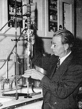
Royal Rife Revisited: Abstract -- In this paper the authors relate information obtained from over three years of work spent researching and reconstructing a working replica of Royal Rife's original Ray Tube apparatus. A description of Rife's discovery of the pleomorphic nature of microorganisms is given along with details of how this led him to invent a revolutionary non-invasive pathogen devitilization technique. Also discussed with limited detail was Rife's development of his extraordinary microscopes. The authors attempt to give the reader new insights into this exciting, readily available technology. Finally, the authors describe the design and fabrication of a complete and working beam tube system, constructed with antique and surplus electronic components. This paper attempts to provide enough information so that all can understand what it was Rife was attempting to accomplish, how Rife's machines worked, and how similar machines might be manufactured today. Also, a list of the original known Mortal Oscillatory Rates (MORs) is provided. No claims for the use of this device in healing human subjects are made. Introduction -- Royal Raymond Rife (1888 - 1971) was an accomplished scientist and microbiologist who developed an optical microscope that could provide magnifications and resolutions heretofore unheard of. He was able, through special quartz optics and a creative optical heterodyning technique, to obtain these resolutions even though they surpassed the theoretical limits of ordinary visible light microscopy. Theoretically, the wavelength of the source illumination is the limiting factor in achievable resolution. It is not possible to image something smaller than the wavelength of the microscope's light source. That is why electron microscopes (with far shorter wavelength) can be used today to image extremely small objects. The major difference between visible light and electron microscopy is that, by its nature, electron microscopes destroy the microorganisms while viewing them. Rife's major advantage was that he could observe them in their natural state. His most powerful instrument is said to be the Universal Microscope which had a magnification of 61,000X and a resolution of 30,000 diameters. Compare this with today's state-of-the-art light source microscopes which are limited to approximately 5,000 diameters. Rife began his work with the microscopes in the early 1920's and it was from these original developments that he would make many of his revolutionary discoveries. It is argued that Rife was the first person to empirically prove that virus and bacteria are pleomorphic forms. Pleomorphism is the phenomenon by which one distinct life form mutates into another. Rife basically classified pathogenic bacteria into 10 individual groups. Rife demonstrated that any organism within its group could be transformed morphologically into any other organism within the 10 groups by carefully altering the media in which it was cultured. Of course this discovery contradicts modern microbiology which teaches that a bacteria's morphology is fixed and unchangeable. Rife also discovered techniques for successfully culturing cancer virus. This virus he identified as BX and it was noted that the viruses refracted a purplish red color with a monochromatic beam under his microscope. In fact, Rife discovered that each organism depending on its state would refract unique spectra and have distinct coloration. By the late 1920's and early 30's, Rife had discovered that by irradiating these pathogenic microorganisms with specific frequencies known as MORs for Mortal Oscillatory Rates, he could cause them to devitalize either by interrupting normal cytologic function or by inducing them to mutate into a non-pathogenic form. The instrumentation involved in this irradiation process has been the subject of a great deal of controversy over the past 50 years. After researching books, films, articles and notes, the authors have concluded that Rife irradiated his pathogenic entities with a modulated radio frequency produced by a sophisticated RF plasma discharge. Rife utilized a radio frequency generator that produced between 100 and 150 Watts of power with a carrier frequency between 3.1 Mhz and 35 Mhz. The output from the generator was connected through suitable impedance matching circuitry to a plasma discharge tube with one or more noble gases. It is believed that Helium was the primary gas used although many researchers cite Argon or an Argon mix as the choice ingredient. Further, Rife utilized a standard dial-type vacuum tube audio frequency generator as the modulation source for his radio frequency transmitter. The modulation signal was a square wave and it is assumed he chose this waveform because of its high harmonic content and broad spectral contributions. Rife obtained the original MORs through a painstaking method of tuning the dial of the audio frequency generator while observing the sample pathogen under his microscope. When a frequency was discovered that demonstrated the ability to devitilize a particular microorganism, its dial position was duly noted and marked. The actual frequencies were determined later after the experimental trials. By the mid 1950s the verified original MOR frequency list included 15 different bacteria and viruses. Regardless of what other researchers have said, the authors believe that these 15 frequencies represented the complete list. The following is a listing of these known MORs as compiled by Dr. Robert P. Stafford, M.D. a physician who worked with an original Rife machine from 1957 to 1963: Microorganism Frequency in Hertz Dr. Stafford who is still living in Dayton, Ohio, independently verified some of Rife's work. Dr. Stafford conducted a rat study with the assistance of Dr. Robert Zipf, M.D., who at the time was the Director of Medical Research at Miami Valley Hospital and in addition, the Montgomery County Coroner. Chloroleukemic Sprague-Dawley rats were utilized in the experiment. Although it is beyond the scope of this paper to discuss the study results in detail, the encapsulated summary is as follows: Ten suckling rats were injected with standard doses of rat leukemic whole blood. Of the seven rats which were inoculated and treated with the Rife equipment, three survived without symptom. Four of these rats died. However, the average time to death was 50.5 days as compared to the group of three non-treated rats which had an average death time of 43.6 days. In addition, all the non-treated rats died. Clearly, even with the four 'failed-cures', in the group of treated rats the Chloroma was favorably impressed. Details of the Original Tube -- In the "Cancer Cure that Worked", Barry Lynes includes a quote penned by Rife himself, which describes the principles of the original Ray Tubes. Also, photographs of original tubes can be found in "The Rife Way III" by Mark Simpson. Figure 1 details the component arrangement of the early units (prior to the formation of Beam Ray Corp.). As described in the introduction, a standard frequency generator (a) was utilized as the square wave modulation source. The individual MORs were selected for broadcast via this instrument. The function generator was connected to the radio frequency generator (b). The square wave signal from the frequency generator was employed in a screen-grid modulation arrangement with the final amplifier of the radio frequency generator. The RF generator incorporated circuitry bearing resemblance to a standard radio transmitter and had a single stage crystal controlled oscillator connected to a class-C amplifier stage. Most likely, the oscillator used either a 6AG7 or 6V6 vacuum tube, while the output was obtained from a pair of standard RF transmitter tubes. RCA designed 807's were introduced in the later models (1940's). The output from the amplifier was connected to an impedance matching network (c) designed to maximize the transfer of RF power from the generator to the gas plasma. As with any transmitter, maximum power transfer occurs when the impedance of the generator is the same as the impedance of the load. The major problem with matching to a plasma is that the plasma's impedance is dynamic and highly non-linear. Rife must have been faced with a formidable challenge having to address the many variables involved in controlling an RF plasma. The original plasma tubes were modified X-ray tubes (d). The tube elements were left intact, however, he tube was refilled with one or more noble gases. When the plasma discharge occurred, the tube glowed purplish blue as attested to by Dr. Stafford, John Crane (Rife's research assistant from 1950 - 1971) and others. Later, Rife had several tubes fabricated from scratch. It is not known precisely how many of this genus of tube were manufactured. Although records are difficult to accurately verify, it is generally thought that patients were treated every third day by exposing them to each frequency on the list for three to four minutes. Normally, only a subset of frequencies were used, a popular list being 728, 784, 880, 2008, and 2128 Hertz. While the modulation frequencies were being broadcast to the patient, the discharge tube was moved over the body of the subject. It was determined that this kind of motion greatly enhanced the experimental effects. Also, an additional improvement was obtained when the MOR frequencies were dithered +/- 10 Hertz. Some believe that in combination with the MOR rates, the RF generator was gated on and off at a 4 hertz rate. Early film footage of the Rife tube in operation does tend to support this conclusion as the tube is seen to flash repetitively. Building a working Beam Tube -- To date, the authors have constructed a total of four Rife units. Two units were fabricated around modified Heathkit transmitters and delivered 100 and 250 Watts respectively. Two additional units were designed from scratch, one utilizing a single 6DQ6 tube as both an oscillator and amplifier. This unit produces approximately 50 Watts of input power. The second and favorite system utilizes a 6AG7 crystal controlled oscillator stage driving a single 807 amplifier. This unit produces about 75 Watts and is ideal for work with RF plasmas. The 807 is an excellent RF amplifier tube in that it is extremely forgiving and will deliver the full rated 75 Watts output with only 220 milliwatts of grid drive. Each of the units employs a solid state modulator in the cathode circuit of the amplifier stage. This cathode arrangement acheives 100% modulation more readily than the screen-grid type originally employed by Rife. The schematic for this unit can be seen in Figure 2. RF Driver, click here to view larger format (121K) The most difficult part of the reconstruction effort was obtaining the glasswork for the plasma discharge tube. Several models were constructed using estimates obtained from descriptions and photographs of original tubes. In addition, physical dimensions were taken from several X-ray tubes designed in the 1940's. Tube fabrication was fabulously expensive and required many tedious hours spent measuring the effective impedance of various gases under varying pressures and power levels. A tremendous amount of time was spent researching plasma chemistry in an attempt to understand the physics behind radio frequency plasma formation and control. If fabricating a replica tube sounds cost prohibitive, please note that a suitable discharge tube can be obtained from a neon sign manufacturer. This less expensive option is highly recommended for a novice experimenter. Have a local glass shop make a 16-19" straight tube and ask them to fill it with argon or helium. This type or discharge tube will operate quite reliably and require no periodic maintenance. Detailed discussions on RF plasmas are beyond the scope of this paper, so it shall be left to the reader to pursue further information on the subject. Most technical libraries will have several informative selections pertaining to RF plasma chemistry. Referring to Figure 2, starting with the oscillator stage, a 6AG7 tube was chosen because its plate load resistance is close to the input impedance of the grid of the 807. This serves to maximize power transfer from the oscillator to the amplifier stage. These impedances can be calculated by using equations and tube data found in older Amateur Radio Handbooks. The 807 requires -45 volts on the grid along with 3.5 milliamps of grid drive for proper excitation. The screen voltage nominally is 300 and the plate can be run at 600 d.c. volts. The driving characteristics of the 807 present an input impedance of approximately 11,000 ohms. A standard tuned-plate tuned-grid crystal oscillator circuit was taken from the handbook along with the recommended component values. The crystal used was cut for 16.0 Mhz. The screen grid of the 6AG7 was fed through a variable 20,000 ohm potentiometer so that the necessary 3.5 milliamps of the grid drive could be carefully adjusted. This control prevents the oscillator from overdriving the amplifier tube. The output from this stage was coupled to the 807 with a simple series capacitor. A fixed regulated supply was chosen over a grid-leak configuration for the grid bias circuit because if the oscillator were to fail, the 807 could be severely damaged. The fixed supply serves as a protective bias for the amplifier. The plasma discharge tube presents an approximate impedance of 2000 ohms at 16.0 Mhz. With the 807 operating with 600 volts on the plate and 150 miliamps of plate current, the output impedance is 4000 ohms, therefore, the discharge tube is tapped down on the output tank coil enabling it to present a higher impedance to the amplifier tube via the transformer action of the circuit. Since the load impedance is matched at 4000 ohms, and the desired loaded Q of the circuit is approximately 15, the tank inductor should exhibit a reactance of 266 ohms at the carrier frequency. This equates to an inductance of 2.65 microhenrys at the 16.0 Mhz operating frequency. A variable capacitor with a range of 5 to 50 picofarads was chosen to resonate with this coil. Since the discharge tube is highly non-linear and has reactive components contributing to its overall impedance, proper adjustment of the variable capacitor allows almost complete cancellation of these reactive components. When the tank circuit is tuned to resonance, the load impedance appears to be purely resistive. This enables the power to be completely transferred from the amplifier to the discharge and be dissipated as radiant energy. Operation and adjustment of the RF driver is fairly simple. Power is first applied to the filaments of the tubes and the tubes are allowed to warm up for several minutes. After the tubes have reached nominal operating temperature, with the amplifier cathode circuit open, high voltage is applied. The drive tuning and drive level of the oscillator stage are adjusted to produce 3.5 milliamps of grid drive for the 807. Next, the cathode circuit is closed and the plate tank capacitor is adjusted to obtain the brightest discharge, which should also require the least amount of plate current, since the circuit will be at resonance with all reactances cancelled. The tap for the discharge tube on the tank coil is adjusted to produce the desired 150 milliamps of d.c. plate current. Modulation can now be accomplished by connecting a suitable square wave function generator to the modulation input. The square waves should have a peak to peak level between 10 and 15 volts. A model B&K-3011 function generator will serve nicely in this capacity. Conclusion -- Now that the Rife Beam Tube technology is fairly well understood and can be duplicated with a high degree of confidence, the next logical step might be to repeat the laboratory studies originally performed by Dr. Stafford et al. Also, a concerted effort should be undertaken towards recreating a working Rife type microscope. Much has been written recently about dark-field microscopy and advances made in Canada by Gaston Naessens and others. Their efforts need to be supported if this technology is ever to be fully realized. This paper did not discuss the "Rife Machines" recently popularized by a number of commercial equipment manufactures. This type of unit normally incorporates either hand-held electrodes or foot plates. It is believed that these devices emerged in the 1950s and are more a product of John Crane than Royal Rife. Rife's involvement in the development of this genre of machine is at best unclear. Although the authors have heard of positive experimental results obtained with the electrode units, they feel that the original Beam Tube system invented by Rife himself offers the best hope as a viable treatment modality. The authors would raise a caution to any individual utilizing any device like this as part of a therapeutic regimen. For additional information on reconstructing a working Rife Beam Tube, contact the authors through the Phoenix chapter of the USPA. [Update: The authors are preparing a second paper, and may be contacted at]: Open Sesame References: "The Cancer Cure that Worked", Lynes, Barry, (1987), pp. 72-73. "The Rife Way III", Simpson, Mark A., (1991), pp. 2-13. "The New Microscopes", Seidel, R.E. and Winter, M. Elizabeth, Journal of the Franklin Institute, February, 1944. "Observations on Bacillus and Typhosus in its Filtrable State", Kendall, Arthur and Rife, Royal, California and Western Medicine, December 1931. "Observations with the Rife Microscope of Filter-Passing Forms of Microorganisms", Science, August 26, 1932. "Techniques and Applications of Plasma Chemistry", Hollahan and Bell, (1974), pp. 393-399. "The Radio Amateur's Handbook", American Radio Relay League, Newington, Conn. "RCA Power Circuits -- D.C. to Microwave", Radio Corporation of America, Harrison, N.J. "Care and Feeding of Power Grid Tubes", Eimac Division of Varian, San Carlos, Ca., (1967). "RCA Transmitting Tubes", Radio Corporation of America, Harrison, N.J. "The Semiconductor Data Library", Motorola Semiconductor Products, Inc., Pheonix, Ariz.
RETURN TO INDEX
Original Rife frequencies Tetanus 120 (An acute, often fatal infectious disease caused by the anaerobic, spore forming bacillus Clostridium tetani.) Syphilis 660 (Caused by Treponema pallidum, a helical, tightly coiled, motile spirochete, a helical to sinusoidal bacterium. Mechanisms of T. pallidum pathogenesis are poorly understood. Existing diagnostic tests for syphilus are sub-optimal, and no vaccine against T. pallidum is available. The subspecies of T. pallidum cause syphilis, yaws, nonvenereal endemic syphilis or pinta.) Gonorrhoea 712 (A gram-negative bacteria, Neisseria gonorrhoea, causes this sexual disease and primarily affects columnar epithelium in genital mucosal surfaces of the urethra, accessory ducts and gland, as well as endocervix. If contaminated fingers rub the eye then conjunctivitis can result.) Staphlococcus 728 (Genus of nonmotile gram-positive bacteria that are found in clusters and that produce important exotoxins. Staphylococcus aureus (Staphylococcus pyogenes) is pyogenic, an opportunistic pathogen and responsible for a range of infections including severe sepsis, pneumonia, endocarditis and soft tissue infections.) Pneumococcus 776 (Gram-positive pyogenic organisms about 1m diameter, usually encapsulated, closely related to streptococcus, associated with diseases of the lung. Pneumococcus is an important cause of serious infections in the first three months of life. These infections are unlikely to be prevented by the currently available infant immunization strategies. One potential approach to prevention of pneumococcal disease in early infancy is immunization of pregnant women.) Streptococcus 880 (A genus of bacteria that are gram-positive cocci, often occurring in chains of varying length. Some pathogenic species produce exotoxins. In man, streptococcal species are responsible for numerous infections such as scarlet fever, tonsillitis, erysipelas (skin infection), endocarditis, rheumatic fever, glomerulonephritis, impetigo, pneumonia, meningitis, pharyngitis, lymphadenitis and wound infections. Streptococcus pneumoniae is the main culprit in lobar-pneumonia and broncho-pneumonia. Streptococcus pneumoniae has been known for more than 100 years as the most important bacterial pathogen of the respiratory tract in adults and children. In recent years, the pneumococcus has begun to exhibit increasing resistance to antimicrobial agents.) Typhoid Bacteria 712 (Typhoid is an infectious febrile illness usually spread by contamination of food, milk or water supplies with bacteria Salmonella typhi. This is not to be confused with Salmonella typhimurium which is the cause of salmonella food poisoning.) Typhoid Virus 1862 Bacillus Coli Rod Form 800 (Most probably Escherichia coli, the archetypal bacterium for biochemists, used very extensively in experimental work. A rod shaped gram-negative bacillus (0.5 x 3-5 m) abundant in the large intestine (colon) of mammals at about .1% of the total. E. coli, along with other species of bacteria, provide us with Vitamin K and B-complex vitamins. But a rare strain of this bacteria, E. coli O157:H7, is responsible for food poisoning which causes bleeding of the intestines which can be fatal.) Bacillus Coli Virus 1552 Tuberculosis Rod Form 803 (Tuberculosis, an infectious bacterial disease caused by Mycobacterium tuberculosis, is characterized by inflammatory infiltrations, formation of tubercles (solid elevations of skin or mucous membranes), tissue death, abscesses, formation of fibrous tissue, and calcification of tissue. Infection is transmitted from infected people, cows, or contaminated milk. Presently the worlds leading killer. It usually occurs as pneumonia, but TB can also occur in the brain, back, knee, lymph nodes, or other organs and bones.) Tuberculosis Virus 1552 Sarcoma cancer (all forms) 2008 (A form of cancer that arises in the supportive tissues such as bone, cartilage, fat or muscle.) Carcinoma cancer (all forms) 2128 (A malignant new growth that arises from epithelium, found in skin or, more commonly, the lining of body organs, for example: breast, prostate, lung, stomach or bowel. Carcinomas tend to infiltrate into adjacent tissue and spread (metastasize) to distant organs, for example: to bone, liver, lung or the brain.) Streptothrix 784 (A genus of bacilli occurring of the form of long, smooth and apparently branched threads, either straight or twisted. Streptothrix is a synonym for Actinomyces israelii. This species is a gram-positive, cast-forming, non–acid-fast, non–spore-forming anaerobic bacillus that is difficult to isolate and identify. Its filamentous growth and mycelialike colonies have a striking resemblance to fungi. They are soil organisms. It can cause the eye diseases Canaliculitis and Keratitis.) Also, frequencies for leprosy, polio, cholera, actinomycosis, glanders, bubonic plague, anthrax, influenza, herpes, cataracts, glaucoma, colitis, sinus, ulcers were discovered by Rife but we don't have any direct records from him on these frequencies. -------------------------------------------------------------------------------- (Definitions in parentheses are mostly from the on-line medical dictionary at http://cancerweb.ncl.ac.uk/omd/index.html and other medical web sites.) Rife stated they had narrowed the actual distinct number of groups of
pathogenic bacteria to 10. In his 1953 book, Rife commented on this: Rife contended certain conclusions escaped earlier researchers simply because they lacked the evidence of their eyes in seeing these forms develop from a single entity: pleomorphism. They require a power of magnification and resolution beyond the typical 2,000 power instrument. Rife's work suggested that the wide array of disease bacterium were merely differentiation phases in a life-cyle of an as of yet undetermined entity. Researcher Gaston Naessens has verified many of Rife's findings, and has delineated 16 phases of change of what Rife called the premodal identity, which Naessens calls "somatids". The Rife frequency instrument kills the "normal" carcinoma cancer cell by rupturing the thousands of BX cancer viruses they contain and thereby dumping the BX cancer virus contents into the cancer cell cytoplasm. This BX cancer virus as Rife named it in 1931 is not a virus by the normal standard usage of the term today. Rife based his definition on the fact that the BX cancer virus could pass through the finest Berkefeld porcelain filter of the time (000 filter). The BX cancer virus is ovoid in shape, .066 microns along the major axis and .05 microns along the minor axis. It is motile, driven by a proton transport flagella the same as its bacterial parent, the E-coli bacteria. When the BX cancer virus is ruptured it spills out its genome, ribosomes, RNA, enzymes, and various proteins. When thousands of these ruptures occur all at once in a carcinoma cancer cell the results are fatal to the cancer cell. A similar situation occurs in the sarcoma cancer cell when the BY cancer viruses are all disintegrated at once. The BY cancer virus is another form of the BX cancer virus which Rife found caused sarcoma cancer after it had been exposed to prolonged ultraviolet light exposure.
The BX was isolated from ten different cases of breast carcinoma by Dr. Royal Raymond Rife at the Rife Research Laboratory in SanDiego (Point Loma), California. It was carried through forty-four transplants on "K" media in all ten instances. The technique used in the isolation of this organism is in brief as
follows; blocks of tissue, taken under the most sterile The BX is a filterable virus, which filters through the W Berkfeld filter.
It is a small ovoid granule, highly plastic, and visible only An inoculating serum was prepared by combining in a mixture, the transplants from the ten original growths with a (?) to 1 dilution of normal saline solution. (The symbol (?) indicates that the text was cropped out in the photocopy.) On Aug. 3, 1933 1/10 cc of the above serum was inoculated into the breasts
of two sets of white rats; one set consisting of On Aug. 28, a set of male rats consisting of the same number was inoculated
as in the females. The same type of epidermal foci (*) Tissue placed in "K" media and run through the original method of technique. It has been demonstrated by experiment that the BX exists in two cycles,
which may be classified as forms A and B. Form A Since experiments show that the Bacillus X in form A exists in malignant
tissue, it is theoretically possible to change its cycle to (note: the frequency listed here is the base frequency that undergoes
modification by the audio frequency that isn't listed here which is believed
to be 2127 or 2008.)
The Forty Year Legacy of Tainted Polio Vaccine In the late 1940's and early 1950's the polio virus was taking a savage toll on the American public. Thousands of children and adults were crippled or killed. In 1955, Jonas Salk performed a medical miracle when he discovered how to mass produce polio vaccine by growing it on the kidneys of rhesus monkeys. While there is no question that thousands were saved from the ravages of polio by the Salk vaccine, by 1960 a problem had surfaced -- a problem which would come back to haunt the nation some forty years later. The complication researchers had isolated in 1960 was a viral contaminate. It seems that when the live polio virus grown on monkey tissues was extracted for vaccine production another virus was extracted as well, SV-40. When this monkey virus was injected into research animals it produced brain cancer. It appears our government didn't wish to create a public panic or discredit the public health service, because instead of recalling the tainted vaccines, it quietly ordered the manufacturers to find a monkey free of SV-40 and continue production. As of 1963, the rhesus monkey had been replaced with the African green monkey for production of a safer polio vaccine, but between the years of 1955 and 1963 as many as 98 million Americans had received doses of live polio virus vaccines tainted with SV-40. Jumping to the early 1990's, Michele Carbone, Assistant Professor of Pathology at Loyola University in Chicago, isolated fragments of the SV-40 virus in human bone cancers and in a particularly nasty form of lung cancer called mesotheliomas. The viral contaminate from the 50s was back to haunt us, and appeared in 33% of the osteosarcoma bone cancers studied, in 40% of other bone cancers, and in 60% of the mesotheliomas lung cancers. Dr. Carbone believed this study could explain why 50% of the current mesotheliomas being treated were no longer occurring in association with their traditional cause of asbestos exposure. Already sounding like a bad science fiction story, the worse news was
yet to follow. An Italian team of researchers from the Institute of Histology
and General Embryology of the University of Ferrara lead by Dr. Fernanda
Martini discovered SV-40's presence in various other tumors. The more this matter is researched the more startling the evidence. Senior epidemiologist at the National Institutes of Health, Dr. Howard Strickler, has plotted a geographic pattern to the cancers associated with SV-40 helping to confirm its link to the tainted vaccine. People who lived in Massachusetts and Illinois who received identified lot numbers of the contaminated vaccine administered in the 1950s are now demonstrating ten times the rate of the osteosarcoma bone tumors as those who received vaccine free of the SV-40 contaminate in other parts of the country. The Food and Drug Administration (FDA) mandates that every American infant and child receive polio vaccinations. While public health officials continue to emphasize how current supplies of the vaccine are safe, Peter Reeve, FDA Virologist, has acknowledged that the administration abandoned independent testing of vaccine purity some fifteen years ago. The job of ensuring safety and purity rests squarely on the shoulders of those manufacturing the vaccines with no federal oversight. Wyeth-Lederle controls the supply of all the oral polio vaccine in this country, and last year's sales totaled some $230 million dollars. Surely there would be no conflict of interest in allowing this corporation to be the sole agent of quality oversight of their own pocketbook? The government may not have paid attention to the quality of these vaccines, but they had formulated a plan for their distribution. Federal vaccination policy advocated the use of live-virus oral polio vaccine (OPV) based on the belief the live virus shed in the body fluids of infants immunized with OPV could immunize others through contact exposure. The Centers for Disease Control (CDC) insisted this was a safe practice, and emphasized that no one previously vaccinated could contract the disease in this manner. The public was never informed of this strategy, however, and no consent was ever obtained from the unknowing participants in this vaccination scheme. One hundred and twenty people, many previously vaccinated, contracted polio as a result of this practice. To add insult to injury in 1994 the World Health Organization proclaimed polio was eliminated from the Western Hemisphere. Insult because for the past seventeen years the only cases of polio occurring in the United States have been caused by the vaccine itself, and injury because this victory will be paid for in blood from the cancers produced by the monkey virus spread with the vaccine. One might ask just how such a thing could happen considering the injectable form of the vaccine (IPV) does not use a live virus and doesn't transmit the disease it is designed to shield us from? Well, Wyeth-Lederle's leading competitor Connaught produces IVP which could explain why Wyeth lobbied so hard against the CDC recommending increased use of IVP. In 1996 the CDC revised its recommendation from four doses of OPV to two doses of IVP followed by two doses of OPV, however, physicians have been instructed to give all four doses as OPV if they desire. The cost of IVP vaccine is $5.40 per dose, whereas OPV costs $2.32 per dose. With the difference in cost favoring the use of OPV, and the current climate of regulating health care costs, clearer guidelines must come from the government if they truly expect to increase the use of the safer IVP vaccine. Well the story of contaminated polio vaccine is not over yet. Microbiologist Howard Urnovitz, Ph.D. provided significant evidence at the Eighth Annual Houston Conference on AIDS that human immunodeficiency virus type 1 (HIV-1) is a monkey hybrid virus which was produced when 320,000 Africans were injected with polio virus contaminated with live simian immunodeficiency virus (SIV) in the late 1950's. Apparently, viral fragments combine easily with other viruses to produce these hybrids called "chimeras." This theory was confirmed by another research team headed by Dr. B. F. Elswood at the University of California in San Francisco. Interestingly enough, when researchers Cecil H. Fox and John Martin applied to the National Institutes of Health for grants to confirm the presence of SIV and simian cyto-megalovirus (SCMV) contaminates in polio vaccines their requests were denied. Dr. Urnovitz may have an explanation as he stated in the Boston Globe, "that almost 100 million Americans were exposed (to SV-40) through a government sponsored program, but for over 30 years, there has been virtually no government effort to see if anyone's been harmed by the exposure." He added, "The government will not fund science that makes it look culpable." Could it be our government, once again, is attempting to avoid a public panic while ignoring the great potential for harm these viruses could inflict. Time will tell. Harvard Medical School professor, Dr. Ronald Desroier points out that taking all known scientific evidence into account that the medical experts' knowledge is limited to "perhaps 2% of existing monkey viruses." Who knows what lethal virus may be discovered in our blood streams forty years from now as a result of good intentions.... References Berleur, M. P., & Cordier, S. (1995). The Role of Chemical, Physical,
or Viral Exposures and Health Factors in Neurocarcinogenesis: Implications
for Epidemiologic Studies of Brain Tumors. Bookchin, D., & Schumaker, J. (1997). Tainted Polio Vaccine Still
Carries Its Threat 40 Years Later. Carbone, M., et al. (1996). SV-40 Like Sequences in Human Bone Tumors. Elswood, B. F., & Stricker, R. B. (1995). Polio Vaccines and the
Origin of AIDS. Fisher, B. L. (1997). Workshop on Simian Virus 40: A Possible Human Polyomavirus. National Vaccine Information Center, January 27, On-line at http://www.909shot.com/polio197.htm>http://www.909shot.com/polio197.htm. Krieg, P., Amtmann E, Jonas, D., Fischer, H., Zang, K., & Sauer
G. (1981). Episomal Simian Virus 40 Genomes in Human Brain Tumors. Lednicky, J. A., Garcea, R. L., Bergsagel, D. J., & Butel, J. S.
(1995). Natural Simian Virus 40 Strains are Present in Human choroid
Plexus and Ependymoma tumors. Martini, F., et al. (1995). Human Brain Tumors and Simian Virus 40. Martini, F., et al. (1996). SV-40 Early Region and Large T Antigen in
Human Brain Tumors, Peripheral Blood Cells, and Sperm Fluids From Healthy
Individuals. Pass, H. I., Kennedy, R. C., & Carbone, M. (1996). Evidence for
and Implications of SV-40 Like Sequences in Human Mesotheliomas. Rock, A. (1996). The Lethal Dangers of the Billion Dollar Vaccine Business. Tognon, M., et al. (1996). Large T Antigen Coding Sequences of Two DNA
Tumor Viruses, BK and SV-40, and Nonrandom Chromosome Changes in Two
Glioblastoma Cell Lines.
By Gary Wade [This is a revised issue, which takes into account some new information received on Rife's frequency instrument. Figures 2 and 3 have been revised and the old text commented on.] During the 1920's and 30's, Dr. Royal Raymond Rife produced some rather astounding accomplishments in medicine and biology. First, he invented a new kind of optical microscope. This microscope could be used to observe viruses in live cells and tissue culture.1,2 Rife built five of these microscopes. Rife never published the plans to his microscope and to this day those in the scientific establishment not familiar with Rife's work wrongly claim that it is generally impossible to see or identify a virus with any optical microscope. Rife's second great accomplishment was to invent a variable frequency flashing light source which could kill bacteria, rickettsias, protozoa, fungi, and viruses. By 1939, Dr. Rife had both identified the microbes and the light flashing rates (frequency) required to kill these microbes, which were associated with fifty-two major diseases, including carcinoma and sarcoma cancers.2,3 In this article we will describe what Rife's frequency instrument was and how it could destroy a microbe without harming the host/patient. Figure 1 shows a schematic view of the Rife frequency instrument in operation treating a patient. The frequency instrument consisted of an old style X-ray tube, which had been back filled with helium gas at very low pressure and had a current flow through the tube driven by a sine wave voltage oscillator. When the voltage polarity across the X-ray tube was such that the hot tungsten cathode was at a negative voltage as compared to the metal plate anode, an electron current would flow from the cathode to the anode. This electron current would collide with the helium atoms exciting them and thereby generating light. Since the polarity conditions for current flow through the X-ray tube were only met on one half the sine wave voltage cycle there was one pulse of light produced for each complete sine wave voltage oscillation cycle (see Figures 2
and 3)
Another way to say this is, that if a million cycles per second of sine wave voltage is applied across the X-ray tube, the X-ray tube will produce one million pulses of light per second. As a practical example, Rife found that the common carcinoma breast cancer, (which is now reaching epidemic proportions among women), was killed by a light pulse of 11,780,000 light pulses per second.4,5 [Note:Probably should be 23,560,000 pulses per second. It now seems that the helium gas pressure (probably .003 milli torr) and voltage (Vrms=928 volts) difference between the cathode and plate were such that electric current flows in both directions in the X-ray tube. This would give twice the light pulses per second than previously believed.gw] For the currently trained biologist and medical researcher, all of the above statements about Rife's work and accomplishments are suspect at best. The reason for this is that they have a very limited knowledge of physics and no knowledge of Rife's research results. For example, they do not know that Rife isolated a viral sized, spore like, motile form of the E-coli bacteria that when exposed to prolonged ultraviolet light became a virulent carcinogen, which invariably caused carcinoma cancer when injected into lab animals. The key to getting Rife's work and accomplishments into general medical and biological use is [to] end to this ignorance about how and why Rife's frequency instrument worked to kill microbes. To that end I will now describe and illustrate how the Rife frequency instrument can destroy a virus. I will illustrate how a specific virus can be destroyed by a specific frequency of ultra sound. This ultra sound is generated by the Rife frequency instrument. Note that light carries linear momentum and that when the pulse of light from the Rife frequency instrument is absorbed by the patient's skin layer that skin layer must recoil in the direction of light flow to conserve linear momentum. When the light pulse has ended, the skin relaxes back toward its non light pulse exposure position. In other words, periodic light pulses generate periodic pressure pulses in the patient's skin layer which travel into the patient's body. The Rife frequency instrument converts the patient's entire exposed skin surface into an ultra sound transducer for the generation of ultra sound. Even though the efficiency of ultra sound production is exceedingly low by this method, it is still adequate to kill microbes. As a practical example, Rife would treat his cancer patients using his frequency instrument for three minutes of exposure once every three days. Usually his "terminally" ill cancer patients would be cancer free in about thirty such treatments or less, as was verified in the 1934, 1935, and 1937 test clinical trials carried out by the U.S.C. Medical School Special Medical Research Committee.3 That same committee then suppressed the research results. The reason for the short three minute treatment is to kill off only a thin outer layer of cancer tumor at one time. This allows the body's immune system to remove this layer before the next treatment. The entire cancer tumor could have been killed/destroyed in a single Rife frequency instrument treatment of perhaps one to one and a half hours. However, then the cancer patient would have a large mass or masses of dead cancer tissue in them, which would become a feast for a massive bacterial infection. This bacterial infection could lead to liver and kidney damage and general toxemia. The Rife frequency instrument kills the "normal" carcinoma cancer cell by rupturing the thousands of BX cancer viruses they contain and thereby dumping the BX cancer virus contents into the cancer cell cytoplasm. This BX cancer virus as Rife named it in 1931 is not a virus by the normal standard usage of the term today. Rife based his definition on the fact that the BX cancer virus could pass through the finest Berkefeld porcelain filter of the time (000 filter). The BX cancer virus is ovoid in shape, .066 microns along the major axis and .05 microns along the minor axis. It is motile, driven by a proton transport flagella the same as its bacterial parent, the E-coli bacteria. When the BX cancer virus is ruptured it spills out its genome, ribosomes, RNA, enzymes, and various proteins. When thousands of these ruptures occur all at once in a carcinoma cancer cell the results are fatal to the cancer cell. A similar situation occurs in the sarcoma cancer cell when the BY cancer viruses are all disintegrated at once. The BY cancer virus is another form of the BX cancer virus which Rife found caused sarcoma cancer after it had been exposed to prolonged ultraviolet light exposure. To see how the ultra sound generated by the Rife frequency instrument can destroy a virus we will examine the outer protein coat (capsid) structure of a virus. Most viruses of interest which cause diseases in plants and animals have an icosahedral capsid structure as illustrated in Figure 4A and B. A
specific example of this icosahedral capsid structure is illustrated in Figure 5 . Each dark circle represents a spherical protein molecule clump. When the virus capsid of Figure 5 is folded together as indicated in Figure 4A and B, a simple virus capsid model has been formed. Examination of this capsid model shows a large number of intersecting and overlapping closed rings of protein molecule clumps. These closed rings of periodically spaced protein clumps are illustrated in Figure 6A and B
In classical physics when studying standing wave phenomenon the periodically spaced protein clumps, as illustrated in Figure 6A and B, are known as the mass beads on a string problem with circular boundary conditions. Figures 7A, B and C
illustrate this classical physics problem. Figures 8A and B illustrate one of the standing wave motion modes which the closed periodically spaced protein clump rings of Figures 6A and B can sustain. Figure 8A shows a ten member protein clump ring linearized for ease of graphing wave motion displacement of the center of the protein clumps from their equilibrium position. Figure 8B
shows the most stressful oscillation mode for the ten member protein clump ring. In this oscillation mode, adjacent protein molecule clumps are always going in opposite directions and therefore putting maximum stress on where they are bonded together. If this oscillation mode is raised to a high enough displacement amplitude the ring will rupture. If enough rings are ruptured, the virus capsid disintegrates. The Rife frequency instrument when set to the frequency which corresponds to the most stressful oscillation mode for the virus of interest, as illustrated in Figure 8B, will destroy that virus capsid coat and therefore destroy the virus. The common virus capsid coat was chosen to show how the Rife frequency instrument can destroy a microbe which have closed on themselves periodically spaced protein clump structures. Bacteria, protozoa, rickettsias, and fungi all have these closed on themselves periodically spaced protein clump structures in their outer structure, which makes them susceptible to destruction by the Rife frequency instrument. Advancements in electron technology have made a much more efficient and vastly more powerful replacement for the Rife frequency instrument. Namely, the piezo-electric transducer, driven by an appropriate signal function generator as can be purchased in any electronic test equipment store. In our present circumstances where antibiotic resistant bacteria are about to become rampant, anti-viral drugs are largely still just a bio-tech dream, and the war on cancer has been a dismal failure for the cancer patient, but not for the so-called cancer researcher. It is long since time for Rife's 1930's work to be implemented. References The New Microscopes, by R.E. Seidel, M.D. and M.Elizabeth Winter, Journal
of the Franklin Institute, Vol.237, Feb. 1944. Chronic Fatigue Alternative Treatment THE HOUSTON POST, March 20, 1991, section A-10 Your Health/Medicine Dr. Resh, an accomplished cell biologist, is exploring how RNA tumor viruses cause normal cells to become cancerous. She is focusing on fatty acylated oncoproteins, which bind to the cell membrane. Her work on membrane-bound viral proteins has expanded to include the Gag proteins of HIV, and she is now studying how these proteins bind to the cell membrane and form a virus particle.
LONDON, Feb 20 (Reuters) - Viruses that cause the common cold could hold important clues to ways of tackling cancer, Scottish scientists said Tuesday. About one-sixth of the 200 different types of cancer, including cervical
cancer and some kinds of leukemia, are triggered by viruses but how they
do it is still a mystery.
Studies have long shown a link between certain strains of the human
papillomavirus and cervical cancer. The sexually transmitted human papillomaviruses
(HPV) include those that cause genital warts and viruses that elicit
no symptoms at all. Cervical cancer appears more common in women with
AIDS. AIDS patients also have a higher rate of precancerous growth on
the cervix, called dysplasia. http://www.usc.edu/hsc/info/pr/ccr/w98/first.html
Click here to Receive our Amazing free newsletters
|

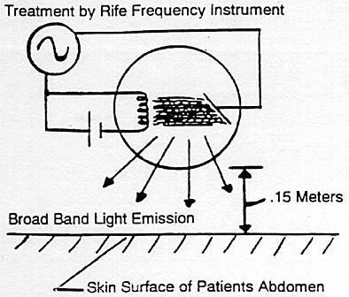
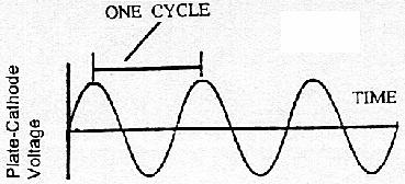
 .
. 
 .
. ,
,