RIFE CRANE Rife machines report on Electron Therapy |

 |
|
 |
|
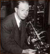
ELECTRON THERAPY RESEARCH John Crane Page 1 This report on Electron Therapy is primarily concerned with the detection and cure of cancer and related diseases. It is presented for research only. It is a matter of record that, while orthodox medical and surgical methods have had limited success with cancer discovered in the early stage, they have had little or no success with treatment of the more advanced cases. The cause of cancer is still listed in some of the current medical textbooks as unknown. Since research to determine the cause and effect a cure of cancer is being carried out in many and various directions, it seems reasonable to explore every possible avenue toward a true understanding of the cause of cancer and hence, its proper treatment This report concerns itself with the discovery of a virus causing cancer and a method of treatment which excited considerable interest among reputable doctors and laboratory specialists as long ago as 1934. This theory has stood up under hundreds of controlled laboratory tests and has been applied to treat human cancer in dozens of cases. Rife Virus Microscope Institute, in conjunction with various doctors throughout the United States and Canada, is at the present time (1960-ed.) engaged in carrying out a controlled series of tests in order to re-establish the finding of Dr. Royal R. Rife in his life study of the cause and cure of cancer. Dr. Royal Raymond Rife first conceived the idea of the Frequency Instrument in conjunction with his work in developing the Rife Universal Microscope. Since the determining of the cancer requires the high power of the Rife Microscope, a brief description of the construction and theory of operation of the Rife Microscope will be included in this report. THE UNIVERSAL MICROSCOPE Dr. Royal R. Rife over a period of 30 years designed and built in his own laboratory five microscopes of power and resolution far beyond the so-called law of optical physics. In their power magnification these instruments vary from 9,000 to 50,000 times, far beyond the limits of the standard research instrument. The commercial microscope being manufactured today is inadequate for the observation of filterable viruses of disease (as these minute, live, living entities are less that 1/20 of one micron in dimension). Thus the need for a device which would carry us farther into this important field of endeavor. We will describe in some detail the most powerful of these microscopes, known as the Universal Microscope. The Universal Microscope, which is the largest and most powerful of the light microscopes, developed in 1933, consists of 5,682 parts. Page 2 This microscope derives its name from its adaptability to all fields of microscopical work. The microscope is fully equipped with separate substage condenser units for transmitted and mono chromatic beam, darkfield, polarized, and slit-ultra illumination, and includes a special device for crystallography. The entire optical system of lenses and prisms as well as the illuminating units are made of block-crystal quartz. The illuminating unit used for the examination of the filterable form of disease organisms contains 14 lenses and prisms, three of which are in the hi-intensity incandescent lamp, four in the Risley prism, and seven in the achromatic condenser system. Two circular, wedge-shaped prisms are suspended between the source of light and the specimen being examined. The two prisms are used for changing the angle of incidence of the light passing through the specimen being examined. When the light passes through these prisms, it is divided or split into two beams, one of which is refracted to such an extent that it is reflected to the side of the prism while the second beam is permitted to pass through the prism and illuminate the specimen owing to its chemical constituents. The mounting arrangement on the Universal Microscope permits each of the two prisms to be rotated in opposite directions by a vernier control throughout 360 degrees. This vernier adjustment permits bending the transmitted beam of light at variable angles of incidence while, at the same time, a small portion of the spectrum is projected into the axis of the microscope owing to the chemical constituents of the microorganism. The vernier adjustment permits only a small portion of the spectrum to be visible at any one time, but it is possible to select any portion from one end of the spectrum to the other. When that portion of the spectrum is reached where both the organism and the color band vibrate in exact accord, a definite characteristic spectrum is emitted by the organism. PRINCIPLE OF PARALLEL RAYS In the case of the filter-passing form of the Bacillus Typhosus, a turquoise blue color is emitted and the plane of polarization deviated plus 4.8 degrees. The predominating chemical constituents of the organism are next ascertained after which the quartz prisms are adjusted by means of the vernier control to minus 4.8 degrees so the opposite angle of refraction may be obtained. A monochromatic beam of light corresponding exactly to the frequency of the organism is then passed through the specimen along with the direct transmitted light. This beam permits the observer to view the organism stained in its true chemical color and reveals its own individual structure in a field which is brilliant with light. The rays of light refracted by the specimen enter the objective lens and are carried up the tube in parallel rays through twenty-one light bends to the ocular lens. A tolerance of less that one wave length of visible light is permitted in the core beam of illumination. Iii the standard optical microscope, the light rays tend to converge as they rise higher and finally cross each other, arriving at the ocular lens, separated by a considerable distance. This microscope derives its name from its adaptability to all fields of microscopical work. The microscope is fully equipped with separate substage condenser units for transmitted and mono chromatic beam, darkfield, polarized, and slit-ultra illumination, and includes a special device for crystallography. The entire optical system of lenses and prisms as well as the illuminating units are made of block-crystal quartz. The illuminating unit used for the examination of the filterable form of disease organisms contains 14 lenses and prisms, three of which are in the hi-intensity incandescent lamp, four in the Risley prism, and seven in the achromatic condenser system. Two circular, wedge-shaped prisms are suspended between the source of light and the specimen being examined. The two prisms are used for changing the angle of incidence of the light passing through the specimen being examined. When the light passes through these prisms, it is divided or split into two beams, one of which is refracted to such an extent that it is reflected to the side of the prism while the second beam is permitted to pass through the prism and illuminate the specimen owing to its chemical constituents. The mounting arrangement on the Universal Microscope permits each of the two prisms to be rotated in opposite directions by a vernier control throughout 360 degrees. This vernier adjustment permits bending the transmitted beam of light at variable angles of incidence while, at the same time, a small portion of the spectrum is projected into the axis of the microscope owing to the chemical constituents of the microorganism. The vernier adjustment permits only a small portion of the spectrum to be visible at any one time, but it is possible to select any portion from one end of the spectrum to the other. When that portion of the spectrum is reached where both the organism and the color band vibrate in exact accord, a definite characteristic spectrum is emitted by the organism. PRINCIPLE OF PARALLEL RAYS In the case of the filter-passing form of the Bacillus Typhosus, a turquoise blue color is emitted and the plane of polarization deviated plus 4.8 degrees. The predominating chemical constituents of the organism are next ascertained after which the quartz prisms are adjusted by means of the vernier control to minus 4.8 degrees so the opposite angle of refraction may be obtained. A monochromatic beam of light corresponding exactly to the frequency of the organism is then passed through the specimen along with the direct transmitted light. This beam permits the observer to view the organism stained in its true chemical color and reveals its own individual structure in a field which is brilliant with light. The rays of light refracted by the specimen enter the objective lens and are carried up the tube in parallel rays through twenty-one light bends to the ocular lens. A tolerance of less that one wave length of visible light is permitted in the core beam of illumination. Iii the standard optical microscope, the light rays tend to converge as they rise higher and finally cross each other, arriving at the ocular lens, separated by a considerable distance.
Page 3 These prisms, located in the tube, are adjusted and held in alignment by micrometer? screws in special tracks made of magnelium a metal having the closest expansion coefficient of any metal to quartz. These prisms are separated by a distance of only 30 millimeters. Thus, the greatest distance that the image in the Universal Microscope is projected through any one media, either quartz or air, is 30 millimeters instead of the 160 to 190 millimeters employed in the air-filled type of the ordinary microscope. It is this principle of parallel rays used in the Universal Microscope and the resultant shortening of projection distance between any two blocks or prisms plus the fact that objective lenses can thus be substitutes for oculars, (these oculars being three matched pairs of lO-mm, 7-mm and 4-mm objectives in short mounts) which make possible not only the unusually high magnification and resolution but which serve to eliminate virtually all chromatic and spherical aberration. The universal stage is a double rotating stage graduated through 360 degrees in quarter-minute arc divisions. The upper segment carries the mechanical stage having a movement of 40 degrees, and the body assembly which can move horizontally over the condenser and provide an angular tilt of 40 degrees + or -. The microscope stands 24 inches high and weighs 200 pounds. The base is composed of cast nickel-steel plate, accurately surfaced and equipped with three leveling screws and two spirit levels set at 90 degrees. The course adjustment, a clock thread screw with 40 threads to the inch, slides in a 11/4 dovetail which gibs directly onto the pillar post. The stage, in conjunction with a hydraulic lift, acts as a lever in operating the fine adjustment. A 6-guage screw, having 100 threads to the inch, is worked through a gland into a hollow glycerine-filled post, the glycerine being displaced and replaced at will as the screw is turned clockwise or counterclockwise allowing a 5 to 1 ratio on the lead screw. This hydraulic action assures complete absence of drag or inertia. The fine adjustment being 700 times wore sensitive than the ordinary microscopes, requires a length of time from ten minutes to one-half hour to focus. This time at first glance seems a disadvantage, but it is felt that, for the overall results obtained, the time required is only a slight inconvenience compared to the many years' research and the actual results obtained in isolating and looking upon disease-causing organisms in their true form (to be continued) A 45 minute video (VHS) of Royal Raymond Rife in his laboratory is available from BSRF and this tape shows the inflicting of cancer in laboratory mice, their cure with Rife's ray tube frequency instruments. It is transferred from a 16mm film and is narrated by John Crane. This film is proof that the cure for cancer (one of many) has been covered up! John Crane can be contacted at 4246 Pepper Drive; San Diego, CA 92105. He is dedicated to preserving and promoting the work of Royal Raymond Rife and the Rife Virus microscope Institute. ELECTR0N THERAPY RESEARCH John Crane Page 1
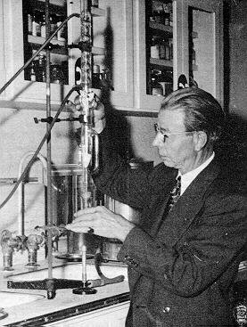
This report on Electron Therapy is primarily concerned with the detection and cure of cancer and related diseases. It is presented for research only. This is part two or two parts. HISTORY OF THE FREQUENCY INSTRUMENTS Dr. Rife's discovery of the various chemical makeup's of different organisms led him to believe that each kind of organism was electrical in nature and should have a resonant frequency determined by each type of chemical combination. His first attempts consisted of a series of unsuccessful experiments to destroy them, with such rays as infrared and ultra-violet. He next turned his attention to radio frequencies. It was his belief that somewhere in the spectrum were frequencies which would resonate with the vibratory rates of disease germs. He believed that if such a frequency could be found and applied, the rays would be fatal to the organism. The first radio frequency instrument was designed and built in 1920. The instrument covered a range of 15 meters to 27,000 meters and was extremely complicated to operate. At that time Dr. Rife was working on the elimination of the Tubercle Bacillus virus. Since the frequency which would affect the virus was unknown, Dr. Rife proceeded by trial and error. Dr. Rife and his assistants made test after test and trial after trial until at last initial success was found. Guinea pigs inoculated with the Tubercle Bacilli and subjected to the frequency instrument at one particular frequency resulted in the organism being killed. At this time a problem even more difficult to solve became apparent. Even though the organism had been killed by the resonance of the proper frequencies, in several cases the inoculated guinea pigs died of toxic poisoning. Three years were spent in determining the answer to this problem. Knowing of the early work of Vaughn with the poison molecule of tuberculosis, he suspected a virus form of the organism was killing his animals. Subsequent studies have demonstrated that this is exactly what had happened. Dr. Rife was then faced with the problem of devising a technique to obtain the virus in pure form, unmixed with contaminating organisms. That Dr. Rife has managed to determine the proper frequency for treatment of this disease and also the treatment of cancer as well as devising the techniques for obtaining the virus of both organisms is a tribute to his remarkable versatility and persistence. "We have been able to devitalize the bacillus and the virus of tuberculosis with radio waves since 1930" Rife said, "but we could not use the treatment until we had done more work to make it possible to use it on man. After being subjected to the waves, the organisms will not grow in culture flasks and cause no disease when injected into guinea pigs." Destruction of the organism by the rays is described as being similar to the phenomenon of a combination of transmitted electronic energy and the coordinate resonance of critical frequencies. This is likened to fragile glass which is shattered by a sustained musical note which is tuned to the resonant pitch. Rife has now demonstrated that the frequency instrument has the power of killing germs that cause tuberculosis and cancer, without harm to human tissue.
Page 2 Development of the Rife Ray to the point where it can be used on beings without harm to human tissue is now a proven fact. Scores of tubercular patients have been treated in private practice and have now recovered. Most cases respond within a period of one to two months and the disease is quickly rendered non-infectious. The isolation of the cancer virus was an accomplishment in which Dr. Rife took a great deal of pride. In 1931 he discovered the transformation of the cancer virus and the successful treatment for cancer by actual observation with the Universal Microscope while applying the frequency instrument treatment. The major portion of the cancer tests of the tumors used in the initial tests was procured from the Paradise Valley Sanitarium in National City, California. The pathology of these tumors was checked through their laboratory as malignant. The methods and principles that were used in this procedure were as follows: An unulcerated breast mass that was checked for malignancy by their laboratory and ourselves came to our laboratory from the paradise Valley Sanitarium. The experiments were carried out in our point Loma Laboratory, then known as the Rife Research Laboratory. A test tube containing' a sample from the unulcerated breast mass was sealed and placed in an Argon gas-filled loop with a two-inch water vacuum and activated with 5,000 volts; the test tube was then incubated for 24 hours. Upon examination of the solution in the test tube, it was found to be teeming with cancer of "B X" virus, which was the most highly motile and the smallest in size of any of the viruses previously isolated. When examined under the Rife Virus Microscope, these B X or cancer viruses refracted a purplish red color with the monochromatic beam. The method of inoculation of experimental animals with B X, the virus of cancer, is as follows: The animal is first shaved and sterilized with alcohol and iodine solution at the point of inoculation and placed under partial anesthesia. This avoids subjecting the anima3 to shock. An extra long,' very small needle is used. The needle is filled with sterile petroleum jelly and a hypodermic is then filled with the inoculum and the needle placed on the syringe. The needle is inserted no less than 30 mm from the point of inoculation under the epidermis. The point of inoculation is in most cases the mammary gland for the reason that the B X involved was recovered from an unulcerated human breast mass. In 3 to 4 days a lesion appears in the thyroid area. The cause of this is unknown, but the lesion recedes and heals over and a growth starts in the mammary gland of the experimental animal. These growths or tumors have exceeded the weight of the experimental animal in many cases. The tumor is surgically removed and the B X is again recovered in all cases. An important factor and check is to make at least 10 transplants from the initial isolation of B X. These transplants are made at 24 hour intervals into the original "K" media. This increases the virulence and speeds the growth of the tumor. With these experiments that have been repeated on over 100 experimental animals, we are convinced that this method definitely proves the virulence and pathology of B X virus.
Page 3 THE TREATMENT "B X" OR CANCER (B X was called Bacillus "X" by Dr. Rife.) The actual cure of cancer in experimental animals occurs with the use of our frequency instrument. To attain these astounding results, a long and tedious process is started to determine the precise setting of the frequency instrument that is the mortal oscillatory rate of this virus. When the setting is found, it is repeated 10 consecutive times after the frequency instrument has been placed back to the same setting before a specific frequency is recorded. These results are observed under the high power of the Universal Microscope and when the mortal oscillatory rate is reached, the B X form5 appear to "blow up" or disintegrate in the field. The inoculated animals are then subjected to the same frequency to determine if the effect is the same on the B X virus in the tissues of the experimental animals. The results are precisely identical with experimental animals as with the pure culture slides; these successful tests were conducted over 400 times with experimental animals before any attempt was made to use this frequency on human cases of carcinoma and sarcoma. The first clinical work 'on cancer was completed under the supervision of Dr. Milbank Johnson, M.D., which was set up under a Special Medical Research Committee of the University of Southern California. Sixteen cases were treated at the clinic for many types of malignancy. After three months, 14 of these so-called hopeless cases were signed off as clinically cured by the staff of five medical doctors and Dr. Alvin G. Foord, M.D., Pathologist for the group, according to Dr. Royal R. Rife. The treatments consisted of three-minutes duration using the frequency instrument which was set on the mortal oscillatory rate for B X or cancer (at 3-day intervals). It was found that the elapsed time between treatments attains better results than the cases treated daily. This gives the lymphatic system an opportunity to absorb and cast off a toxic condition which is produced by the devitalized dead particles of the' B X virus. No rise of body temperature was percept-able in any of these cases above normal during or after the frequency instrument treatment. No special diets were used in any of this clinical work, but we sincerely believe that a proper diet compiled for the individual would be of benefit. THE DETERMINATION AND DIAGNOSIS OF CANCER We can determine in over 90% of the cases of persons having carcinoma by the examination of a blood smear (with the technique heretofore explained) in 30 minutes. We have also found that 'in many types of epithelioma that the carcinoma tissue carries no conductivity with a pendulum galvanometer which enables us to outline and determine the location of a tumor without the use of X-ray photographs. It has also been determined that any case of malignancy treated with either X-ray
Page 4 All pathogenic diseases can be eliminated with preventative treatments using the frequency instruments. We find, in 98% of carcinomous individuals in the monocytes of their blood, granular forms. This is one type of organism we find in blood from carcinomous individuals as Dr. Rife has stated, "Malignancy is a blood disease and this is one type or one stage of this granular form which can be made into carcinoma by growth on "K" media. This is one form of malignancy and from the cryptomyces pleomorphia long, Rife can transfer any of these stages back into a B X and produce a tumor from which can be recovered a true B X or cancer virus. (This article was written over a quarter of a century ago. John Crane can still be contacted concerning the work of Royal R. Rife and the Rife Virus Microscope Institute. Contact: John Crane, 4246 Pepper Drive, San Diego, CA 921O5 - USA. Phone # (619) 281-0278)
September-October 1986 JBR
|